• Climate, Environment & Health
  • Materials

Innovation projects - drug testing, cancer research, anti-MRSA agents & hair transplantation


INNOVATIONS PROJECTS

Body-on-a-chip combines “mini-organs” in a small space, for example to perform drug tests. The chip shown contains the intestine, liver, kidney as well as the blood-brain barrier. Other organs, such as the skin, lungs or heart, are also already in production. An expansion of the chip to ten organs is planned.

PEOPLE ON THE CHIP

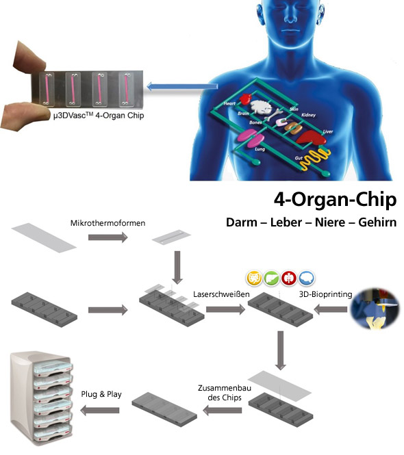
Drug testing on humans repeatedly results in adverse health effects, sometimes even fatal. Animal experiments, on the other hand, are ethically questionable and their results are not always transferable to the human body. Miniaturized human 3D tissues and organs made from different cells, so-called body-on-a-chip, can already provide indications of of drug tolerability in humans in the preclinical phase, thus minimizing the risk in the clinical phases. There is a need for in vitro systems in which supplying blood vessels can be grown in combination with 3D tissues and which meet the physical and mechanical demands of a blood vessel system. Today, there are few microfluidic chip systems, which however consist of angular channels and do not represent the curved environment of a blood vessel or the flow profile of the blood vessel. Prof. Schepers' research group was able to develop a three-dimensional microfluidic blood vessel system consisting of round, porous and branched channels through microthermoforming, onto which miniaturized 3D organs can be 3D bioprinted from human cells. Resulting low-cost disposable products can reduce the cost of pharmaceutical research and minimize the number of animal experiments.

WITH LIGHT AGAINST CANCER

Despite advances in cancer treatment, there are still problems with many chemotherapeutics, such as drug resistance or serious side effects due to the simultaneous killing of stem cells and proliferating tissue. Likewise, it remains a challenge with radiation therapy to avoid damaging surrounding healthy tissue. Photodynamic therapy is an innovative treatment method for cancer based on light-inducible active substances, so-called photosensitizers. The photosensitizer is injected into the patient’s bloodstream and is initially completely harmless to the body. Only when irradiated with light of a certain wavelength does the photosensitizer develop its toxic effect: Local exposure of the tumour results in the formation of reactive oxygen species that destroy the tumors.

However, conventional photosensitizers such as titanium dioxide are not suitable for the treatment of deep-lying tumors, as they are activated with UV light, which has a low penetration depth in the human body and is cell damaging. Photosensitizers, which are activated with longer-wave light, are therefore of great interest for tumor therapy. The research group has developed light-sensitive β-tin tungstate nanoparticles, which, in contrast, are very stable under physiological conditions. Since they are activated with visible light, deeper tumors can also be treated.

FLUORENSCENT SPRAY AGAINST MELANOMA

Malignant melanoma, the black skin cancer, is one of the most dangerous types of skin cancer. If detected in the advanced stage, there is little chance of recovery. If a malignant melanoma is detected early, there is a good chance of recovery. Early detection is mainly performed by surgical removal and histological examination of degenerated liver patches. This method is complex and allows only random examinations of patients.

One of Prof. Scheper’s product ideas is based on the development of a contact gel or a spray for the whole body treatment and the subsequent detection of degenerated skin areas using a commercially available and inexpensive UV lamp that stimulates the fluorescence of the dye and makes the degenerated liver spots visible. Any unintaken substance can be removed by washing it off beforehand. The idea is based on a fluorescent substance synthesized by the research group, which is selectively absorbed only by cells of the malignant melanoma. Screens on human cells do not show uptake in healthy skin cells or in other cells or tumors.

The picture shows the frequency of detection of MSRA in human blood. The group headed by Professor Schepers is researching drugs against the widespread hospital germ.

ANTI-MRSA ACTIVE SUBSTANCES

In Germany, the number of infected patients is around one million per year, with the number of deaths rising to up to 40,000. The reason for this is an infection with one of the three most common hospital germs MRSA, ESBL or the intestinal germ VRE. Multi-resistant germs have adapted over time to the presence of antibiotics. The cells develop strategies to disable their mechanisms of action and then react only poorly or no longer at all to the active ingredients. Multi-resistant germs are found globally, especially in hospitals and nursing homes. There are increasingly fewer treatment options available for multidrug-resistant germs, such as Salmonella or Staphylococcus aureus (MRSA). At present, therefore, it is impossible to contain the formation and spread of these so-called super germs by conventional means.

Prof. Scheper’s team has discovered a new class of compounds based on fat-soluble peptide-like structures that show very good efficacy against MRSA. Unlike conventional antiobiotics, the peptidomimetics do not attack the bacterial metabolism but destroy the bacterial cell membrane, causing the MRSA to dry out. This kills and eliminates the bacteria. Resistance does not develop.

In a research collaboration, Prof. Schepers has developed an alternative to painful hair transplantation. This is based on genetic reproduction of the hair roots and on a biodegradable foil, which is attached to the scalp and then painlessly dissolved with hair roots.

3D HAIR PRINTING AND HAIR TRANSPLANTATION

In Germany alone, around ten million men suffer from hair loss and about one in three women are affected. Usually, the only remedy is a hair transplant. The surgeon removes a strip of skin from the crown of hair at the back of the head. Several thousand hair follicles are located on this skin lobe, which is now broken down into small grafts. The individual hair follicles are transplanted into small holes in the scalp, which the surgeon either cuts, stings or burns with a CO2 laser. About 3000 individual hairs are often transplanted. This procedure is very painful and cannot be performed in patients with little own hair in the wreath.

In collaboration with a group from the Institute of Technical Chemistry, Professor Schepers has developed a procedure that avoides a surgical procedure and allows the insertion of several thousand hairs at a time. In this procedure, a few hair roots are removed from the patient's hair crown by a biopsy at the family doctor's office. Several thousand hair roots are then multiplied from two to three follicles. In order to place these follicles on the scalp in a simple process, the cells are printed on a special polymer foil with small hair-root-like recesses using a newly developed 3D laser printing process. The edges of the recesses are chemically modified so that they act like a velcro tape to which the hair root cells can adhere. Since it is a foil material that is biodegraded by a special enzyme, several thousand follicles can now be transplanted in one step without surgery.

After detachment of the epidermis by fruit acid peeling, as it is done in cosmetic treatment of aging signs, the foils are glued on large areas with fibrin glue. After 48 hours the skin is regenerated and the hair roots are ingrown. Within a short time, the foil decomposes into FDA (Food and Drug Registration) approved products.

Diese Seite nutzt Website-Tracking-Technologien von Dritten, um ihre Dienste anzubieten. Ich bin damit einverstanden und kann meine Einwilligung jederzeit mit Wirkung für die Zukunft widerrufen oder ändern.

Alle akzeptieren Einstellungen Nur notwendige akzeptierenImpressumDatenschutz