Researchers at KIT discover a new possibility for magnetic resonance tomography (MRT) that could revolutionize the production of magnetic resonance tomograms.
MRT yes, but...
Magnetic resonance tomography is a medical imaging procedure that has been used for many years to identify pathological changes in the body, such as inflammation, signs of wearing or tumors in soft tissue. The procedure does not involve harmful radiation, but a good and clear diagnosis is challenging. For each examination, radiology specialists have to weigh up the quality of the tomograms produced and the reasonably short measurement time of this costly procedure. If the examinations take too long, the energy of the radio waves used can heat up the body cells of the person being treated and cause long-term damage. In addition, not all examinations can be carried out without a contrast agent. In particular, if the examined tissue produces homogeneous gray tones in the image, a contrast agent is required to improve the informative value of the image. The heavy metal gadolinium is often used, which is water-soluble but can be deposited in the body in the long term. Researchers at KIT, together with other cooperation partners from Germany and the USA, have now made discoveries that could revolutionize the current MRT procedure in terms of people's health.
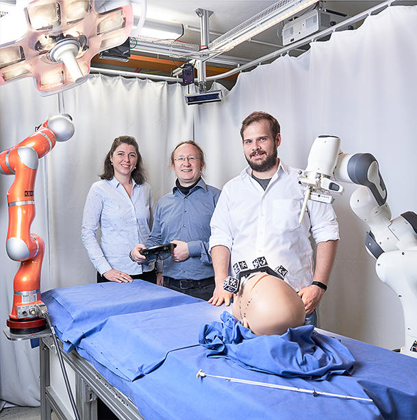
The RASER-MRT approach is based on spontaneous emission without added radio waves. The researchers create the necessary negative spin order using hyperpolarization. (Image: Amadeus Bramsiepe / KIT)
Better results without radio frequency
A conventional MRT image, also known as an MRI, is produced in three steps. The person to be treated is placed lying down in a tube-like MRT device, inside which there is a strong magnetic field. This first aligns the nuclear spins of the hydrogen atoms present in the body, similar to small magnets. In the second step, these are tilted by precisely tuned radio waves. Depending on the composition of the tissue, this brief change generates different signals while the spins realign with the magnetic field. In the third step, the signals are location-encoded using magnetic field gradients, recorded by a computer and converted into a grayscale image.
NUCLEAR SPIN
Atomic nuclei possess a fundamental physical property known as nuclear spin, which represents a kind of magnetic moment.
The novel RASER MRT approach (radio-frequency amplification by stimulated emission of radiation) presented by KIT follows the same three examination steps, but has two significant differences. In the second examination step, the radio wave signal is generated by stimulated emission, similar to a laser. This means that it does not require external, pulsed radio waves. In principle, detection is the same as with conventional MRT images. In order for the spontaneous emission to work in the second step, the researchers generate the necessary negative spin order by hyperpolarization to align the nuclear spins of the molecules. "In application, these can be molecular contrast agents such as pyruvate, for example," explains Dr. Sören Lehmkuhl, research associate in the NMR Microtechnologies for Imaging and Spectroscopy department at the Institute of Microstructure Technology (IMT). The molecular contrast agent is highly magnetized with the help of a catalyst and injected into the person being treated before they are placed in the MRT device. It magnetizes in the opposite direction to the external field and thus generates a significantly higher energetic output state in the body. The signal is thus generated spontaneously without stimulation, as the spins tilt on their own to align themselves along the magnetic field. In addition to measuring without radio frequency excitation, the RASER technology also enables more detailed image results. "As the signal is longer and stronger than with conventional MRT procedures, the resolution achieved is also greater. For radiology specialists, this means that they can make more reliable statements about possible diseases, for example by recognizing from the metabolism whether a cancer therapy is effective or needs to be adjusted," explains Lehmkuhl.
HYPER POLARIZATION
Hyperpolarization in nuclear magnetic resonance describes a process in which a stronger magnetization of the atomic nuclei is achieved than would be possible within the applied magnetic field in thermodynamic equilibrium.
MOLECULAR CONTRAST AGENTS
Molecular contrast agents are particularly promising in the field of NMR spectroscopy, as they not only provide an image, but can also map functions. They are pre-magnetized in order to provide the best possible signal. This allows RASER-MRI procedures to highlight small and low-contrast structures more clearly in the tomogram.
PYRUVATE
Pyruvate is the anion or salt of pyruvic acid, which also occurs in the human body. It is an intermediate product in aerobic and anaerobic metabolism and is produced, for example, when glucose is broken down. Pyruvate is a promising molecular contrast agent and has been used for around 10 years.
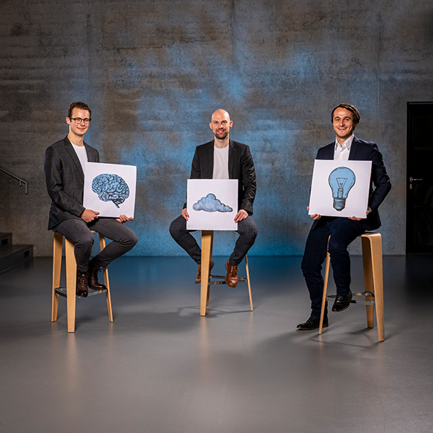
Hyperpolarization is part of the RASER-MRT procedure. The research team hyperpolarizes samples using their own NMR. (Image: Amadeus Bramsiepe / KIT)
From theory to practice
The researchers have worked out the theory and published what they were able to demonstrate in initial proof-of-principle experiments. In the next step, they want to further develop the technology and advance the transition from basic research to application. "We have started to measure RASER on an MRT device. One major challenge is the RASER threshold, which has to be overcome for spontaneous emission. For the spontaneous signal, we need a certain amount of negative polarization and a resonator with the appropriate quality. Applied to our research, this means that we need to further develop both the hyperpolarization technology and the miniaturization of the detectors," says Lehmkuhl, describing the next steps. In addition to the developmental challenges, the approval of molecular contrast agents is also an important issue. "There is currently no approval for hyperpolarized pyruvate in Germany, but approval processes have already been initiated, for example by the University of Heidelberg. The potential of hyperpolarized contrast agents is huge, so I am confident that they will be approved," adds Lehmkuhl.
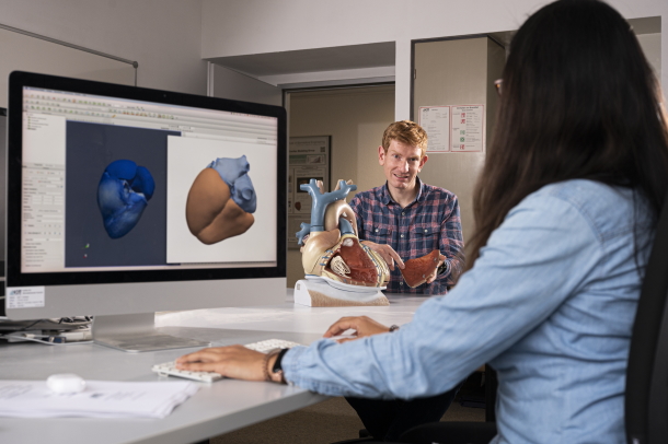
The research team around Dr. Sören Lehmkuhl at the Institute for Microstructure Technology (IMT): Dr. Sören Lehmkuhl, Jakoba Wacker, Simon Fleischer, Elene Aslanikashvilli, Leon Middendorf and Ahmed Hasaneen (from left to right). (Image: Amadeus Bramsiepe / KIT)
Lehmkuhl and his team are cooperating with various partners to overcome the existing hurdles and enable the transfer to society. In the USA, on the other hand, the focus is clearly on the step towards application. Our wide-ranging collaborations enable us to further develop the technology on a holistic level in order to one day bring it into application. Our wish is to implement it in existing MRT devices, as this is feasible and attractive for the market. No radio frequency radiation means less hardware on the MRT scanner and therefore less effort for upgrading and converting existing devices," predicts Lehmkuhl. It will probably be several years before RASER-based MRI is used. However, it is already clear that it could revolutionize the classic MRT procedure and offers a glimmer of hope for faster, more accurate and health-safe diagnostics.
Dr. Sören Lehmkuhl, Research assistant in the department of NMR microtechnologies for imaging and spectroscopy at the Institute of Microstructure Technology (IMT) (Image: Amadeus Bramsiepe / KIT)
The RASER-MRT approach is based on spontaneous emission without added radio waves. The researchers create the necessary negative spin order using hyperpolarization. (Image: Amadeus Bramsiepe / KIT)
Hyperpolarization is part of the RASER-MRT procedure. The research team hyperpolarizes samples using their own NMR. (Image: Amadeus Bramsiepe / KIT)
The research team around Dr. Sören Lehmkuhl at the Institute for Microstructure Technology (IMT): Dr. Sören Lehmkuhl, Jakoba Wacker, Simon Fleischer, Elene Aslanikashvilli, Leon Middendorf and Ahmed Hasaneen (from left to right). (Image: Amadeus Bramsiepe / KIT)
Keyfacts:
GOAL
Investigation and development of an MRT procedure without radio frequency excitation for integration in the research environment and in existing MRT devices
PARTNERS
Research Center Jülich, RWTH Aachen University, Harvard University, Wayne State University, North Carolina State University
AWARDS
Emmy Noether funding, funding by the German Research Foundation (DFG)
Diese Seite nutzt Website-Tracking-Technologien von Dritten, um ihre Dienste anzubieten. Ich bin damit einverstanden und kann meine Einwilligung jederzeit mit Wirkung für die Zukunft widerrufen oder ändern.